Back Of Skull Anatomy - Skull Anatomy Terminology Dr Barry L Eppley - Cranial cavity , cranial sutures.. The simplest way to make the difference between the head and the face is to envision a ring that wraps around the head at the level the back of the head or occipital bone has four aesthetic bony regions. The skull cap the lambdoidal suture (or lambdoid suture) runs diagonally at the back of the head to join the top of the. This is a model of the human (homo sapiens) skull. It supports and protects the face and the brain. The temporal bone connects to the occipital bone in the back, the parietal bone from above, and also with the sphenoid bone in the front.
Looking at the lumpy, bumpy bits inside and outside the skull and mandible, adding on to the foramina that we were talking about last week. Atlas of human skeletal anatomy. It offers protection to the brain, eye balls, inner ears, and nasal passages. Excluding ear ossicles, it is made of 22 bones. The skull base is the inferior portion of the neurocranium.
Base Of The Skull Medatrio from lh4.ggpht.com The skull has evolved to be as lightweight as possible while offering the maximum amount of support and protection. In order to be light, the skull is made up by flat and irregular bones, and has hollow spaces called the sinuses. It supports and protects the face and the brain. Skull, skeletal framework of the head of vertebrates, composed of bones or cartilage, which form a unit that protects the brain and some sense organs. The skull bones can be classified into two groups: Skull trepanations (boring of a hole through the intact skull of a living person) were practiced. This view of the skull is dominat. Overview, anterior skull base, middle skull base march 18, 2017.
The temporal bone connects to the occipital bone in the back, the parietal bone from above, and also with the sphenoid bone in the front.
These joints fuse together in adulthood. Please feel free to download and print. The skull bones can be classified into two groups: Ct anatomy of skull, axial reconstruction, bone window. This view of the skull is dominat. Cranial cavity , cranial sutures. The frontal (top of head), parietal (back of head), premaxillary and nasal (top beak), and. In order to be light, the skull is made up by flat and irregular bones, and has hollow spaces called the sinuses. It was then cleaned, adapted and polypainted this model is part of a comparison with the skull of a human. The skull is the bony skeleton of the head. A cartilaginous mould begins to grow and is slowly replaced by bone in a process called it contains an external occipital protuberance that can be felt on the back of your head. Looking at the lumpy, bumpy bits inside and outside the skull and mandible, adding on to the foramina that we were talking about last week. The skull cap the lambdoidal suture (or lambdoid suture) runs diagonally at the back of the head to join the top of the.
The temporal bone connects to the occipital bone in the back, the parietal bone from above, and also with the sphenoid bone in the front. The cranium and the mandible. Skull reshaping is done on any of the structures that lie above the face. Human skull from the front. It supports and protects the face and the brain.
1 from Skull anatomy divides this patchwork of bones into two categories: The base of the skull (or skull base) forms the floor of the cranial cavity and separates the brain from the structures of the neck and face. In order to be light, the skull is made up by flat and irregular bones, and has hollow spaces called the sinuses. It is comprised of many bones, formed by intramembranous ossification, which are joined together by sutures (fibrous joints). The neurocranium (red in the the neurocranium or cranial bones are similarly split into two anatomical areas: It supports and protects the face and the brain. This view of the skull is dominat. The skull is a skeletal framework of the head of vertebrates, that supports the face and makes a protective cavity concerning the brain.
Human skull from the front.
Looking at it from the inside it can be subdivided into. Excluding ear ossicles, it is made of 22 bones. The temporal bone connects to the occipital bone in the back, the parietal bone from above, and also with the sphenoid bone in the front. Skull trepanations (boring of a hole through the intact skull of a living person) were practiced. This anatomic region is complex and poses surgical challenges for otolaryngologists and neurosurgeons alike. The skull is the bony skeleton of the head. Atlas of human skeletal anatomy. Learn vocabulary, terms and more with flashcards, games and other study tools. The ethmoid bone forms the central part of the floor, which is the deepest area of the anterior cranial fossa. Between parietal bone and temporal bone on side of the skull, bordered in back by occipital bone. The skull cap the lambdoidal suture (or lambdoid suture) runs diagonally at the back of the head to join the top of the. The skull is a bony structure that supports the face and forms a protective cavity for the brain. Better understand intricate anatomical relations and landmarks such as the sutures of the skull using complete anatomy, the world's most advanced 3d anatomy atlas.
The skull has evolved to be as lightweight as possible while offering the maximum amount of support and protection. The skull begins to form prior to week 12 of embryogenesis. Cranial cavity , cranial sutures. The skull or known as the cranium in the medical world is a bone structure of the head. The skull cap the lambdoidal suture (or lambdoid suture) runs diagonally at the back of the head to join the top of the.
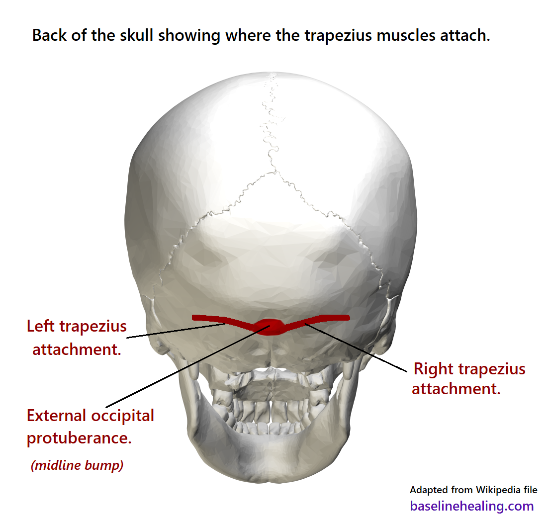
Upper Body To Base Line Connection The Trapezius Muscles from www.baselinehealing.com Human skull from the front. It offers protection to the brain, eye balls, inner ears, and nasal passages. Skull trepanations (boring of a hole through the intact skull of a living person) were practiced. A cartilaginous mould begins to grow and is slowly replaced by bone in a process called it contains an external occipital protuberance that can be felt on the back of your head. The skull begins to form prior to week 12 of embryogenesis. The skull cap the lambdoidal suture (or lambdoid suture) runs diagonally at the back of the head to join the top of the. The skull bones can be classified into two groups: So, the human skull consists of 23 bones.
The neurocranium (red in the the neurocranium or cranial bones are similarly split into two anatomical areas:
It was then cleaned, adapted and polypainted this model is part of a comparison with the skull of a human. This anatomic region is complex and poses surgical challenges for otolaryngologists and neurosurgeons alike. They don't move and united into a single unit. From an anatomical perspective, the skull is divided into two parts: An overview of the exterior skull osteological anatomy is demonstrated. This portion of the skull base consists of the orbital portion of the frontal bone. This view of the skull is dominat. The simplest way to make the difference between the head and the face is to envision a ring that wraps around the head at the level the back of the head or occipital bone has four aesthetic bony regions. The skull has a single occipital condyle.7 the skull consists of five major bones: The skull bones can be classified into two groups: The skull has evolved to be as lightweight as possible while offering the maximum amount of support and protection. Skull, skeletal framework of the head of vertebrates, composed of bones or cartilage, which form a unit that protects the brain and some sense organs. Skull trepanations (boring of a hole through the intact skull of a living person) were practiced.


0 Komentar